
Research Tools

Diffusion Tensor Imaging (DTI)
Diffusion tensor imaging is a type of MRI image that can create images of the white matter of the brain. Each neuron in the brain consists of a cell body, where computations occur, and a long axon that carries the result of that computation to other neurons. The brain's white matter pathways are bundles of axons carrying information from one place to another. Diffusion tensor imaging is based on measuring the natural random drift of water molecules found within brain tissue over the course of a few seconds. Without any physical barriers, water molecules diffuse randomly in every direction, but the tissue in brain structures form barriers that limit the water diffusion in some directions. In white matter, the water diffuse down the length of the axon, but the axon walls limit the diffusion in other directions. A diffusion tensor imaging shows this preferential diffusion. Diffusion tensor images are often color-coded to highlight the spatial orientation of fibers. Specifically, left-right fibers are often shown in red, front-back fibers in green, and up-down fibers in blue. Fibers that are oriented diagonally receive a mixture of these colors, like yellow or purple.

Trascranial Direct Current Stimulation (tDCS)
Transcranial direct current stimulation (tDCS) is a non-invasive technique that applies a very small electrical current (like a 9 Volt battery) to the brain. This slightly enhances or inhibits activity in the parts of the brain affected by the current. tDCS can be used to study which parts of the brain are involved in a particular language or cognitive function, by seeing whether enhancing or inhibiting a part of the brain alters performance on a task. We also study whether tDCS may improve recovery from brain injury by subtly modulating brain activity.

TMS/Electroencephalography (EEG)
EEG measures the electrical activity of the brain. We use EEG during TMS to measure how TMS impacts brain activity. We hope this information will allow us to design better TMS treatments for brain injury.
Behavioral studies
Behavioral studies in healthy subjects typically involve simple computerized tasks. These tasks are used to test hypotheses about the underlying psychological processes involved in particular functions, or to test hypotheses about how the brain performs these functions.

Functional MRI (fMRI)
When a part of the brain is active, the body responds by sending more blood to that area to make sure it can function properly. fMRI examines brain activity by measuring these changes in oxygen in the tiny blood vessels throughout the brain. This can tell us which parts of the brain a person uses to perform particular language or cognitive tasks. Measurements of brain activity at rest allow us to examine which parts of the brain are functionally connected, i.e. those that tend to activate together. In healthy subjects, fMRI can help us understand how the brain usually performs language and cognitive functions. In people with brain injury, fMRI can help us understand how the brain reorganizes to support recovery.
Structural Imaging and Cognitive Neuropsychology
We use MRI to examine brain structure in people with and without brain injuries. MRI uses magnetic fields to provide very detailed pictures of the brain without using any ionizing radiation. This allows us to examine how injuries to specific structures relate to a person’s deficits, and also to examine how treatment or “natural” recovery from injury changes brain structure. We use three different structural MRI techniques: - Lesion Analysis studies the relationship between damaged brain regions and behavior in order to localize function. It is an old technique made much more powerful thanks to accurate imaging! - Voxel Based Morphometry (VBM) is a neuroimaging analysis method that examines tiny differences in brain anatomy between individuals at every location in the brain. This allows us to test whether particular functions or patterns of recovery from injury relate to small anatomical differences in specific brain structures. - Diffusion Tensor Imaging (DTI) allows us to observe the tracks of nerve fibers within the brain. We use DTI to test whether pathways connecting different brain structures are important for language functions or recovery from injury.
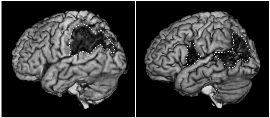
Rendering of left hemisphere lesion (outlined).

Procedure for "skull-stripping" a brain.

Lesion Symptom Mapping
Software for free download
Allows SVR-LSM analyses with flexible handling of covariates including lesion size, permutation-based correction for multiple comparisons, and a graphical interface.
Cite: DeMarco AM and Turkeltaub PE. A multivariate lesion symptom mapping toolbox and examination of lesion-volume biases and correction methods in lesion-symptom mapping. Human Brain Mapping, 2018.